
Shopping Cart
Remove All Your shopping cart is currently empty

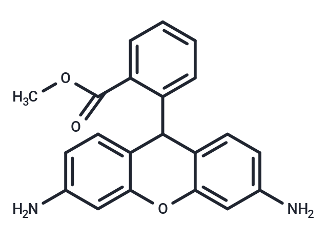
Dihydrorhodamine 123 (DHR 123) is a fluorescent probe (λex=488 nm, λem=525 nm). Dihydrorhodamine 123 is oxidized to the fluorescent reagent Rhodamine 123 in the presence of intracellular reactive oxygen species and is localized in mitochondria.

| Pack Size | Price | USA Stock | Global Stock | Quantity |
|---|---|---|---|---|
| 1 mg | $33 | In Stock | In Stock | |
| 5 mg | $79 | In Stock | In Stock | |
| 10 mg | $112 | In Stock | In Stock | |
| 25 mg | $205 | In Stock | In Stock | |
| 50 mg | $306 | In Stock | In Stock | |
| 100 mg | $455 | - | In Stock |
| Description | Dihydrorhodamine 123 (DHR 123) is a fluorescent probe (λex=488 nm, λem=525 nm). Dihydrorhodamine 123 is oxidized to the fluorescent reagent Rhodamine 123 in the presence of intracellular reactive oxygen species and is localized in mitochondria. |
| In vitro | In the presence of 10 μM Dihydrorhodamine 123 (DHR 123), the stimulation of neutrophil NADPH oxidase with 50 nM phorbol 12-myristate 13-acetate (PMA) increases the rate of rhodamine generation. Similarly, induced HL60 cells exhibit a sustained fluorescence increase after the addition of 50 nM PMA in the presence of 10 μM DHR 123. |
| Cell Research | I. Detection of Reactive Oxygen Species (ROS) 1. Solution preparation: Dissolve DHR 123 in an appropriate solvent (such as DMSO or PBS) at a concentration of typically 1-10 μM. 2. Cell staining: Add DHR 123 solution to cultured cells and incubate at 37°C for typically 30-60 min. 3. Oxidation process: ROS produced in cells oxidize DHR 123, converting it into fluorescent products. 4. Fluorescence measurement: After staining, measure the fluorescence of the oxidation products using a fluorescence spectrophotometer or fluorescence microscope with an excitation wavelength of 488 nm and an emission wavelength of 525 nm. 5. Analysis: Analyze ROS levels in cells by fluorescence intensity. Experiments can be performed in real time or at fixed time points. II. Assessment of mitochondrial function and membrane potential 1. Cell incubation: Incubate cells as described above. 2. Mitochondrial ROS detection: Oxidized DHR 123 will show a fluorescent signal under a fluorescence microscope, indicating the generation of mitochondrial ROS. 3. Fluorescence microscopy observation: Observe the fluorescent signal to see the distribution of ROS in specific areas of the cell (especially mitochondria). III. Flow cytometry for ROS quantitative analysis 1. Cell staining: Add DHR 123 solution to cells as described above. 2. Flow cytometric analysis: After incubation, wash the cells and perform fluorescence analysis using a flow cytometer. The fluorescence intensity is proportional to the ROS level in the sample, which can achieve quantitative analysis. The above information is based on published literature. Experimental procedures should be appropriately modified to meet specific research demands. |
| Synonyms | DHR 123 |
| Molecular Weight | 346.38 |
| Formula | C21H18N2O3 |
| Cas No. | 109244-58-8 |
| Smiles | COC(=O)c1ccccc1C1c2ccc(N)cc2Oc2cc(N)ccc12 |
| Relative Density. | 1.313 g/cm3 (Predicted) |
| Storage | Powder: -20°C for 3 years Shipping with blue ice/Shipping at ambient temperature. | |||||||||||||||||||||||||||||||||||
| Solubility Information | DMSO: 262.5 mg/mL (757.84 mM), Sonication is recommended. | |||||||||||||||||||||||||||||||||||
Solution Preparation Table | ||||||||||||||||||||||||||||||||||||
DMSO
Note : The dilution table applies only to solid products. For liquid products, please calculate the stock solution based on the stated concentration and/or density. | ||||||||||||||||||||||||||||||||||||
 For example, if the intended dosage is 10 mg/kg for animals weighing 20 g , with a dosing volume of 100 μL per animal, and a total of 10 animals are to be administered, using a formulation of
  10% DMSO+ 40% PEG300+ 5% Tween 80+ 45% Saline/PBS/ddH2O , the resulting working solution concentration would be 2 mg/mL.
Dissolve 2 mg of the compound in 100 μL DMSO to obtain a stock solution at a concentration of 20 mg/mL . If the required concentration exceeds the compound's known solubility, please contact us for technical support before proceeding.
1) Add 100 μL of the DMSO stock solution to 400 μL PEG300 and mix thoroughly until the solution becomes clear.
2) Add 50 μL Tween 80 and mix well until fully clarified.
3) Add 450 μL Saline,PBS or ddH2O and mix thoroughly until a homogeneous solution is obtained.

| Size | Quantity | Unit Price | Amount | Operation |
|---|
 Hello! How can I help you today?
 Copyright © 2015-2026 TargetMol Chemicals Inc. All Rights Reserved.
